Doctors order saline infusion images, or SIS - also known as echocardiography, SHG or water ultrasound - as a diagnostic tool for examining internal problems in the uterus. The saline infusion sonogram is a slightly more complex transvaginal ultrasound version, which is the preferred imaging test for assessing female pelvic and reproductive structures because it provides valuable information but is safe and relatively inexpensive during pregnancy. And without the use of ionizing radiation, there may be dangerous side effects. The difference between standard transvaginal ultrasound and uterine ultrasound is that for uterine contrast ultrasound, the uterus is filled with sterile fluid prior to examination; this provides a more pronounced and better intrauterine image than p.
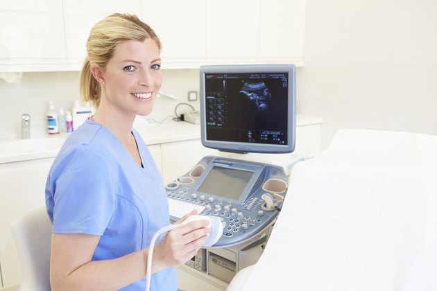 A nurse sits in front of an ultrasound machine. (Photo: monkeybusinessimages / iStock / Getty Images)
 Purpose
When your doctor wants to evaluate the endometrium (medically called the endometrium) to determine Due to abnormal vaginal bleeding, infertility or repeated abortion, saline infusion imaging has greatly increased the information obtained from conventional transvaginal ultrasound. The test can identify several different problems, such as uterine fibroids, endometrial polyps, retained pregnancy products, or congenital anomalies, such as abnormally shaped uterus.
Preparation
Most medical service providers check for cervical infections by doing several cervical cultures before infusion of the sonogram for days or weeks. Your provider passes a small catheter through the cervix to fill the uterus with fluid. In theory, catheters can spread existing infections in the cervix to the uterus, so many providers also require taking prophylactic antibiotics before and after testing. Even with antibiotics, you can get infections or other complications after infusion of saline, but the risk is very small - less than 1%.
Procedure
Do saline - Infusion sonogram, your medical provider first inserts the uterus through the vagina and cervix through a sterile catheter and fills the uterus with sterile fluid. He then positioned the ultrasound probe in the vagina because it was closest to the replica of the effective organ and provided the most accurate image. The probe emits sound waves at frequencies that are inaudible to the human ear. Most transvaginal ultrasound examinations onlyIt takes less than 10 minutes to complete; saline infusion ultrasound usually does not require longer than this.
After the examination
Although you generally do not feel too much pain in the saline infusion image, you may feel some discomfort, often cramps. In addition to the discomfort associated with placing speculum and vaginal ultrasound probes, many women experience painful menstrual cramps as fluid enters and dilates the uterus. Taking over-the-counter painkillers, such as ibuprofen, or even taking an anxiolytic, one to two hours before surgery can help reduce discomfort. After surgery, you usually do not have an activity limit, but mild spasms and hemorrhage usually occur within a few days after the test. If you have a fever, severe or persistent pain, or severe or persistent bleeding after infusion of a saline map, call your health care provider to discuss your symptoms.
Results
Physiological saline infusion imaging is generally considered a good screening for endometrial abnormalities. It can catch abnormalities in the uterus, but it can't distinguish between an abnormality, such as uterine fibroids, and another, such as polyps. In addition, although this test is very reliable in detecting anomalies (if they do exist), false positive readings may occur indicating an error. If your provider normally interprets your test results, subsequent imaging is usually not required. If the saline infusion is abnormal, she would recommend a more specific - usually more invasive - check, such as a hysteroscopy, to verify the results of the saline infusion ultrasound.